Japanese Village, 1921, David Burliuk

Japanese Village, 1921, David Burliuk
Medium: oil,canvas
More Posts from Tools333 and Others
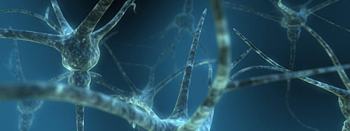
Neural Stem Cells Regenerate Axons in Severe Spinal Cord Injury New relay circuits, formed across sites of complete spinal transaction, result in functional recovery in rats In a study at the University of California, San Diego and VA San Diego Healthcare, researchers were able to regenerate “an astonishing degree” of axonal growth at the site of severe spinal cord injury in rats. Their research revealed that early stage neurons have the ability to survive and extend axons to form new, functional neuronal relays across an injury site in the adult central nervous system (CNS). The study also proved that at least some types of adult CNS axons can overcome a normally inhibitory growth environment to grow over long distances. Importantly, stem cells across species exhibit these properties. The work will be published in the journal Cell on September 14. (For a history of spinal cord repair science and the significance of this latest work, read Ohio State University neuroscientist Phillip Popovich’s review here.) The UC San Diego-led team embedded neural stem cells in a matrix of fibrin (a protein key to blood-clotting that is already used in human neuron procedures), mixed with growth factors to form a gel. The gel was then applied to the injury site in rats with completely severed spinal cords. “Using this method, after six weeks, the number of axons emerging from the injury site exceeded by 200-fold what had ever been seen before,” said Mark Tuszynski, MD, PhD, professor in the UC San Diego Department of Neurosciences and director of the UCSD Center for Neural Repair, who headed the study. “The axons also grew 10 times the length of axons in any previous study and, importantly, the regeneration of these axons resulted in significant functional improvement.” In addition, adult cells above the injury site regenerated into the neural stem cells, establishing a new relay circuit that could be measured electrically. “By stimulating the spinal cord four segments above the injury and recording this electrical stimulation three segments below, we detected new relays across the transaction site,” said Tuszynski. To confirm that the mechanism underlying recovery was due to formation of new relays, when rats recovered, their spinal cords were re-transected above the implant. The rats lost motor function – confirming formation of new relays across the injury. The grafting procedure resulted in significant functional improvement: On a 21-point walking scale, without treatment, the rats score was only 1.5; following the stem cell therapy, it rose to 7 – a score reflecting the animals’ ability to move all joints of affected legs. Results were then replicated using two human stem cell lines, one already in human trials for ALS. “We obtained the exact results using human cells as we had in the rat cells,” said Tuszynski. The study made use of green fluorescent proteins (GFP), a technique that had never before been used to track neural stem cell growth. “By tagging the cells with GFP, we were able to observe the stem cells grow, become neurons and grow axons, showing us the full ability of these cells to grow and make connections with the host neurons,” said first author Paul Lu, PhD, assistant research scientist at UCSD’s Center for Neural Repair. “This is very exciting, because the technology didn’t exist before.” Pictured: Artist’s rendering of neurons
g33k.mk That is so cool! #Repost@natures(@get_repost) ・・・ Growing kidney bean, 25 day timelapse 🌱 Follow @naturesfor more!👈

Lush and Delightful by Marek Boguszak


Ruup Students of Estonian Academy of Arts
“Gigantic wooden megaphones” for the forest inhabit a clearing in Estonia's Pähni Nature Centre. They are part of an acoustic installation meant to amplify the sounds of the landscape, serve as outdoor classrooms or just provide shelter to a weary hiker.
Images Tõnu Tunnel


snow

🚲
(via fuckyeahnativeamericans)


-
 hn-1973 liked this · 5 years ago
 -
 angelamcewen reblogged this · 5 years ago
 -
 tools333 reblogged this · 5 years ago
 -
 queerhistorymajor liked this · 5 years ago
 -
 david-burliuk reblogged this · 5 years ago
