One Of The Most Dangerous Pictures Ever Taken - Elephant’s Foot, Chernobyl. This Is A Photo Of A Now
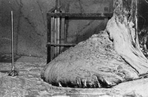
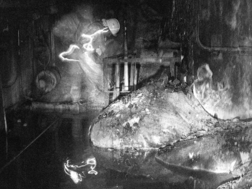
One of the most dangerous pictures ever taken - Elephant’s Foot, Chernobyl. This is a photo of a now dead man next the ‘Elephant’ Foot’ at the Chernobyl power plant.
The image distortions in the photo are created by intense level of radiation almost beyond comprehension. There is no way the person in this photo and the person photographing him could have survived for any more that a few years after being there, even if they quickly ran in, took the photos and ran out again. This photo would be impossible to take today as the rates of radioactive decay are even more extreme now due to a failed military experiment to bomb the reactor core with neuron absorbers. The foot is made up of a small percentage of uranium with the bulk mostly melted sand, concrete and other materials which the molten corium turns into a kind of lava flow. In recent years, it has destroyed a robot which tried to approach it, and the last photos were taken via a mirror mounted to a pole held at the other end of the corridor for a few seconds. It is almost certainly the most dangerous and unstable creation made by humans. These are the effects of exposure: 30 seconds of exposure - dizziness and fatigue a week later 2 minutes of exposure - cells begin to hemorrhage (ruptured blood vessels) 4 minutes - vomiting, diarrhea, and fever 300 seconds - two days to live
More Posts from Science-is-magical and Others


Here are 17 jaw-dropping photos of space that show us just how small we really are:
This photo of the moon and Earth taken from the International Space Station.

A dwarf galaxy, about 11 million light-years away from us.

Earth as seen from the moon in 1968.

A cluster of stars, 20,000 light-years away from Earth.

The first flower grown in the International Space Station, photographed by astronaut Scott Kelly.

Saturn, seen through an infared filter.

These visible “loops” on the surface of the sun can reach up to 15 times the diameter of Earth in height.

The Northen Lights just North of Chicago, viewed from the International Space Station.

The Quintuplet Cluster, located 100 light-years from the center of our galaxy.

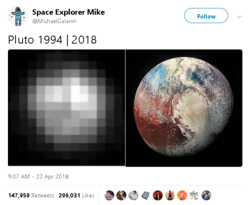
Pluto and one of its moons, Charon.

The Great Pyramids of Giza, seen from space.

Astronaut Bruce McCandless maneuvering, untethered, above Earth in 1984.

Galaxy NGC 6240, 400 million light-years away from Earth.

Palomar 12, a cluster of stars on the outskirts of the Milky Way.

The remnants of an exploded star.

New York City, seen from the International Space Station.

And the remains of a supernova whose explosion may have been seen almost 2,000 years ago by Chinese astronomers.
Follow @the-future-now
Newborn baby brain scans will help scientists track brain development
Scientists have published ground-breaking scans of newborn babies’ brains which researchers from all over the world can download and use to study how the human brain develops.

(Image caption: Diffusion MRI of the developing neonatal brain (Left: Multi-shell high angular resolution diffusion data decomposed into a free water component (greyscale background image) and a directionally resolved brain tissue component shown as rendered surfaces. Middle and right: Visualisation of anatomical connections in the developing brain derived from the brain tissue component.)
The images are part of the Developing Human Connectome Project (dHCP), a collaboration between King’s College London, Imperial College London and the University of Oxford, which will uncover how the brain develops, including the wiring and function of the brain during pregnancy and how this changes after birth. The dHCP researchers are sharing their images and methods online so that other scientists from around the world can use the data in their own research.
Using Magnetic Resonance Imaging (MRI) scanners at Evelina London Children’s Hospital, the team has developed new techniques which enable images of the brains of foetuses and babies to be captured. Researchers have overcome problems caused by babies’ movement and small size, as well as the difficulties in keeping vulnerable infants safe in the MRI scanner, so that they can now produce highly detailed and rich information on brain development.
The project will help scientists to understand how conditions such as autism develop, or how problems in pregnancy affect brain growth.
‘The Developing Human Connectome Project is a major advance in understanding human brain development - it will provide the first map of how the brain’s connections develop, and how this goes wrong in disease,’ said Lead Principal Investigator, Professor David Edwards from King’s College London and Consultant Neonatologist at Evelina London.
The research collaboration is funded by a €15 million Synergy Grant from the European Research Council, and one of the goals of the project is to make sure that the data is shared as widely across the world as possible. Scientists are able to download the images at https://data.developingconnectome.org.
For this project, Professor Jo Hajnal’s team at King’s College London developed new MRI technology specifically designed to provide high resolution scans of newborn and fetal brains.
In addition, a group led by Professor Daniel Rueckert’s at Imperial College London developed new computer programs to analyse the images. 'We have been developing novel approaches that help researchers by automatically analysing the rich and comprehensive MR images that are collected as part of dHCP,’ he explained.
At the University of Oxford, Professor Steve Smith’s team has been developing specific techniques to define where the connections are in the developing brain.
As well as studying more babies, the team at Evelina London are now recruiting pregnant mothers for foetal scanning. Meanwhile, the first release of images today will allow scientists to start to explore these powerful images and begin mapping out the complexities of human brain development in a whole new way.

(Image caption: The above image compares the neural activation patterns between images from the participants’ brains when reading “O eleitor foi ao protesto” (observed) and the computational model’s prediction for “The voter went to the protest” (predicted))
Brain “Reads” Sentences the Same in English and Portuguese
An international research team led by Carnegie Mellon University has found that when the brain “reads” or decodes a sentence in English or Portuguese, its neural activation patterns are the same.
Published in NeuroImage, the study is the first to show that different languages have similar neural signatures for describing events and scenes. By using a machine-learning algorithm, the research team was able to understand the relationship between sentence meaning and brain activation patterns in English and then recognize sentence meaning based on activation patterns in Portuguese. The findings can be used to improve machine translation, brain decoding across languages and, potentially, second language instruction.
“This tells us that, for the most part, the language we happen to learn to speak does not change the organization of the brain,” said Marcel Just, the D.O. Hebb University Professor of Psychology and pioneer in using brain imaging and machine-learning techniques to identify how the brain deciphers thoughts and concepts.
“Semantic information is represented in the same place in the brain and the same pattern of intensities for everyone. Knowing this means that brain to brain or brain to computer interfaces can probably be the same for speakers of all languages,” Just said.
For the study, 15 native Portuguese speakers — eight were bilingual in Portuguese and English — read 60 sentences in Portuguese while in a functional magnetic resonance imaging (fMRI) scanner. A CMU-developed computational model was able to predict which sentences the participants were reading in Portuguese, based only on activation patterns.
The computational model uses a set of 42 concept-level semantic features and six markers of the concepts’ roles in the sentence, such as agent or action, to identify brain activation patterns in English.
With 67 percent accuracy, the model predicted which sentences were read in Portuguese. The resulting brain images showed that the activation patterns for the 60 sentences were in the same brain locations and at similar intensity levels for both English and Portuguese sentences.
Additionally, the results revealed the activation patterns could be grouped into four semantic categories, depending on the sentence’s focus: people, places, actions and feelings. The groupings were very similar across languages, reinforcing the organization of information in the brain is the same regardless of the language in which it is expressed.
“The cross-language prediction model captured the conceptual gist of the described event or state in the sentences, rather than depending on particular language idiosyncrasies. It demonstrated a meta-language prediction capability from neural signals across people, languages and bilingual status,” said Ying Yang, a postdoctoral associate in psychology at CMU and first author of the study.


-
 ik1ru liked this · 4 months ago
 -
 nuanced-nuisance reblogged this · 7 months ago
 -
nuanced-nuisance liked this · 7 months ago
-
 derogatorylt reblogged this · 9 months ago
 -
 theunbearablelightnessofbein liked this · 1 year ago
 -
 massaodanno liked this · 1 year ago
 -
 themightyif liked this · 1 year ago
 -
 doctorjohnsmith liked this · 1 year ago
 -
 gentlemensmuse liked this · 1 year ago
 -
 blast-door liked this · 1 year ago
 -
 symdayruram liked this · 1 year ago
 -
 hermes-stropheus reblogged this · 1 year ago
 -
hermes-stropheus liked this · 1 year ago
-
 intoxicateddaily reblogged this · 2 years ago
 -
 napea liked this · 2 years ago
 -
 emb666 reblogged this · 2 years ago
 -
 swinginreadingfictiondean liked this · 2 years ago
 -
 kiddohc liked this · 2 years ago
 -
 impaled liked this · 2 years ago
 -
 eclectichellmouth reblogged this · 2 years ago
 -
 comeandjudge reblogged this · 2 years ago
 -
 mary-maud reblogged this · 2 years ago
 -
 d4sgl4s liked this · 2 years ago
 -
 burningzipperhumanoidpizza reblogged this · 2 years ago
 -
 clepsidra-so liked this · 2 years ago
 -
 justdees reblogged this · 2 years ago
 -
mary-maud reblogged this · 2 years ago
-
 bs-rottengir reblogged this · 2 years ago
 -
 k4ertu reblogged this · 2 years ago
 -
k4ertu liked this · 2 years ago
-
 mg-dl reblogged this · 2 years ago
 -
 ooscar88 reblogged this · 2 years ago
 -
ooscar88 liked this · 2 years ago
-
 laura-mv reblogged this · 2 years ago
 -
 koulamarababushka liked this · 2 years ago
 -
 nauseabogota liked this · 2 years ago
 -
 musdomus reblogged this · 3 years ago
 -
 nauticalgotham liked this · 3 years ago
 -
 willow-wildflowers reblogged this · 3 years ago
 -
 stairwell-flowers reblogged this · 3 years ago
 -
stairwell-flowers liked this · 3 years ago
