Lachryphagy Is The Term Used To Describe The Behaviour Of Tear Drinking In Nature, Typically In Environments
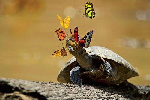
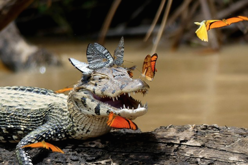
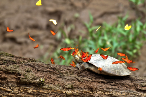

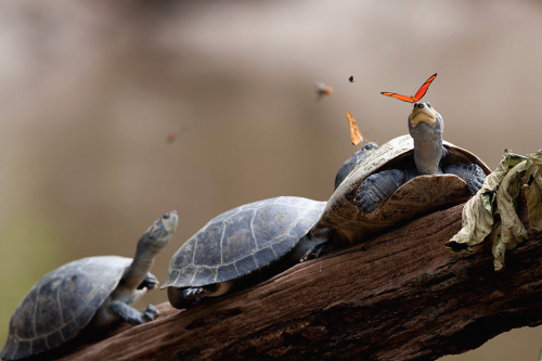
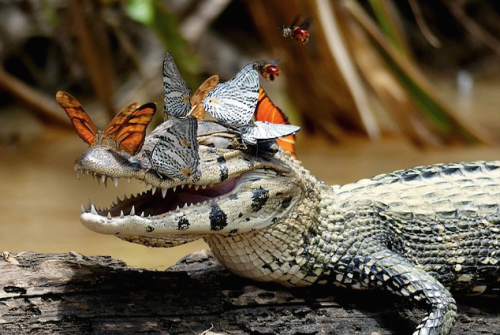
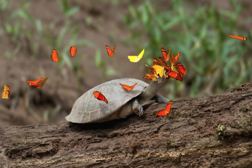
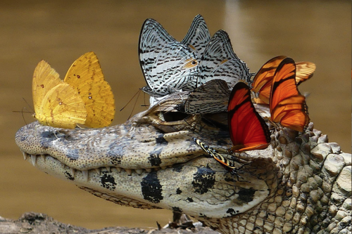
lachryphagy is the term used to describe the behaviour of tear drinking in nature, typically in environments - like the purvian amazon shown here - where sodium and other micronutrients are hard to find.
bees and butterflies need sodium for egg production and metabolic purposes, but their diets of nectar are low in salt. so the orange julia and sulfur yellow butterflies you see here turn to the salty tears of often stationary turtles and caiman.
and though the caiman and turtles seem to receive no reciprocal benefit from the interaction, they’re apparently happy enough to just help out. (x, x, x, x, x, x)
More Posts from Ricardocedillob and Others


Gantz by Hiroya Oku via Ominous - 不吉


BLOOMINGTON, Ind. — With crisp resolution to 100 nanometers, the DeltaVision OMX imaging system is considered one of the world’s finest microscope systems. Upon its arrival to Indiana University Bloomington’s Light Microscopy Imaging Center in 2010, researchers quickly renamed it the “OMG” microscope for the amazing images it produced and for its ability to do super-speed imaging of multiple-labeled proteins in cells.
A metaphase epithelial cell stained for microtubules (red), kinetochores (green) and DNA (blue), was the winning image, submitted by IU, in the 2012 GE Healthcare Life Sciences Cell Imaging Competition. The DNA here is fixed in the process of being moved along the microtubules that form the structure of the spindle.

A researcher was walking through a city market when he came upon a piece of dinosaur tail, encased in amber and preserved for millions of years in all its feathery glory.
Our curator of dinosaurs says it could help settle a debate over how feathers evolved in the first place.
Image of the Week – June 8, 2020
CIL:12375 - http://cellimagelibrary.org/images/12375
Description: Movie showing the dynamics of kinetchore microtubules during meiosis II in primary spermatocytes of the crane-fly Nephrotoma suturalis that were experimentally flattened. Time-lapse polarization microscopy using a Nikon Microphot SA, equipped for liquid crystal polarized light microscopy (LC-PolScope, CRi, Woburn Massachusetts) 60x/1.4 PlanApo oil immersion objective, 1.4 NA oil imm. condenser, with 2.0x zoom lens. Images captured every 15 sec using a QImaging Retigo EXi CCD camera. Raw images were processed using 5-frame algorithm (Shribak and Oldenbourg, 2003). The time series used for the movie is included in this grouped set.
Authors: James R. LaFountain and Rudolf Oldenbourg
Licensing: Public Domain: This image is in the public domain and thus free of any copyright restrictions. However, as is the norm in scientific publishing and as a matter of courtesy, any user should credit the content provider for any public or private use of this image whenever possible.

Tag yourself, I’m lysosome. (Not my picture/content)

Will Turner
-
 buttercupfromthebronx reblogged this · 1 month ago
 -
buttercupfromthebronx liked this · 1 month ago
-
 elektrostantsiya liked this · 1 month ago
 -
 wolf-halls reblogged this · 1 month ago
 -
 bitsofsciencelife reblogged this · 2 months ago
 -
 catfeetpicdealer reblogged this · 2 months ago
 -
 7thgenscot liked this · 2 months ago
 -
 teamliberty reblogged this · 2 months ago
 -
 coolstufftothinkabout reblogged this · 3 months ago
 -
 pharaoh-markaba reblogged this · 4 months ago
 -
 sangxdencre liked this · 4 months ago
 -
 keeniebeenie liked this · 4 months ago
 -
 mon-t-real reblogged this · 4 months ago
 -
 voyagesurlalune reblogged this · 4 months ago
 -
voyagesurlalune liked this · 4 months ago
-
 2nd-me reblogged this · 4 months ago
 -
2nd-me liked this · 4 months ago
-
 jmanning1986 liked this · 5 months ago
 -
 simurguvercinka liked this · 5 months ago
 -
 carr10n-fl0wers liked this · 5 months ago
 -
 abloodygoodmess reblogged this · 5 months ago
 -
 thelochnesscreature reblogged this · 6 months ago
 -
thelochnesscreature liked this · 6 months ago
-
 severeprincesheep reblogged this · 6 months ago
 -
severeprincesheep liked this · 6 months ago
-
 fuckyeahilike reblogged this · 6 months ago
 -
 annita89q56p4bh liked this · 7 months ago
 -
 acrasiomycetes reblogged this · 8 months ago
 -
 whoiisshe liked this · 8 months ago
 -
abloodygoodmess liked this · 8 months ago
-
 itstoodreamy liked this · 8 months ago
 -
 feathereddragonkin liked this · 9 months ago
 -
voyagesurlalune reblogged this · 9 months ago
-
 oh-shush reblogged this · 9 months ago
 -
 tmajik60 liked this · 10 months ago
 -
 warcotuj8746 liked this · 10 months ago
 -
 chryso-poeia reblogged this · 11 months ago
 -
chryso-poeia liked this · 11 months ago
-
 traceamountsoftimetravel reblogged this · 1 year ago
 -
 lolimrandom liked this · 1 year ago
 -
 ppppranger reblogged this · 1 year ago
 -
 dasghboard liked this · 1 year ago
 -
 mngolv reblogged this · 1 year ago
 -
 terracottahearted liked this · 1 year ago
